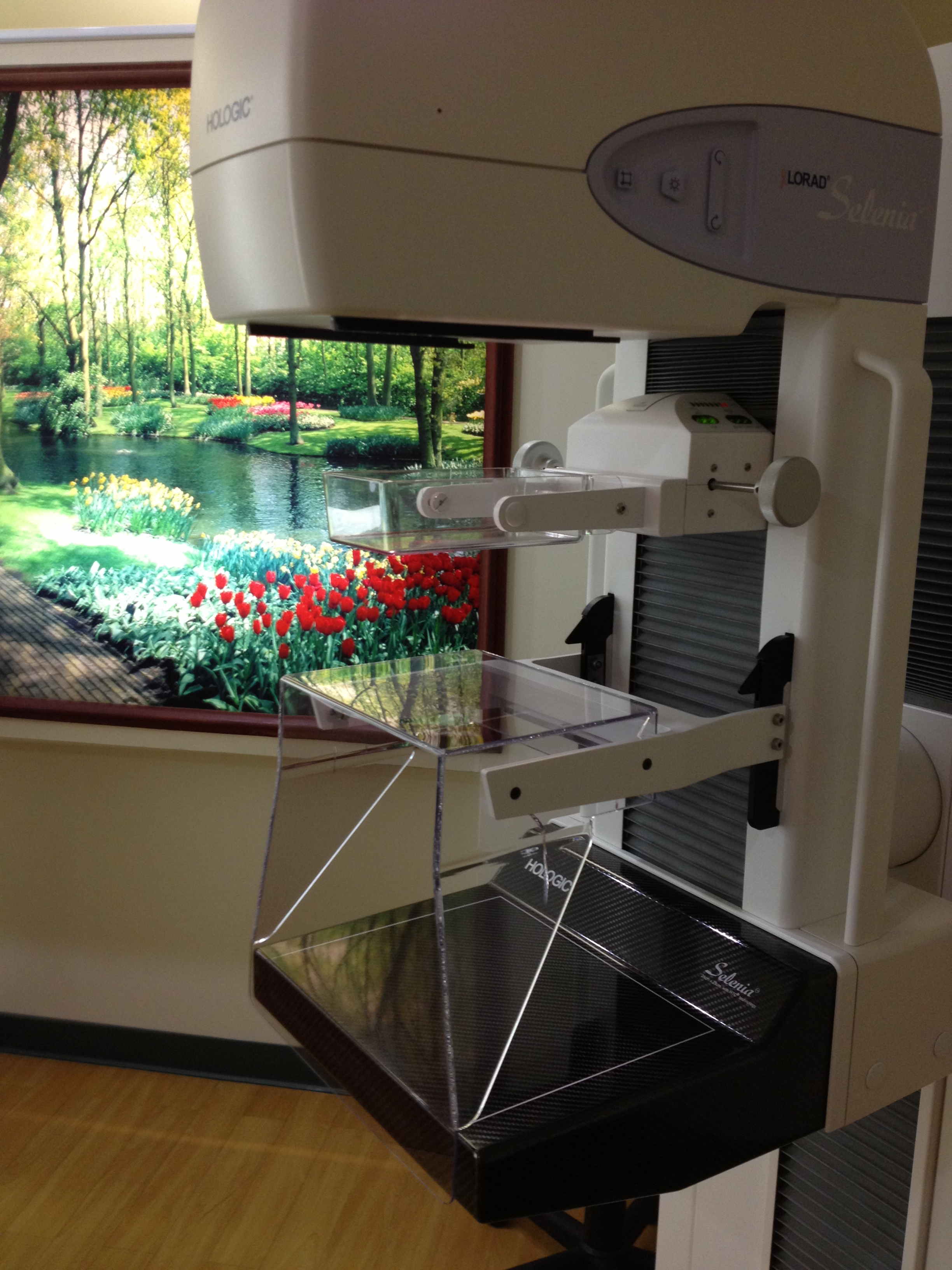
We are the first and only regional facility to offer Hologic’s Selenia Full Field Digital Mammography. Clearly shown to increase the rate of early detection, digital mammography was our choice and should be your choice. Large studies have shown that there was a significant increase in the number of breast cancers detected following the switch from the old-fashioned mammography equipment that uses film to Digital Mammography.  Because of the better detection of breast cancer, all of the best medical centers in the U.S. now use only Digital Mammography. The list includes Harvard University’s Medical Center, UCLA, Cedars Sinai Medical Center, The Mayo Clinic, UCSF, Johns Hopkins, Stanford and basically al top centers. There are still many places that do not use Digital Mammography. But, why not select the best detection available. After all, isn’t the earliest detection possible what we are all after?
Fred S. Vernacchia, M.D., is a prominent researcher in the field of early breast cancer detection who Dr. Vernacchia has authored numerous medical articles, lectured to other radiologists internationally and has served as a director on many non-profit boards. He recently stated, that, based on his experience: “I would certainly encourage patients who are being screened to look for facilities that have digital technology because it is faster and has a higher cancer detection rate.” We agree and hope you’ll make the right choice and select Digital Mammography.
What is Mammography?
Mammography is a specific type of imaging that uses a low-dose x-ray system to examine breasts. A mammography exam, called a mammogram, is used to aid in the early detection and diagnosis of breast diseases in women.
Two recent advances in mammography include digital mammography and computer-aided detection. What do they add?
Digital mammography, also called full-field digital mammography (FFDM), is what we use at Guam radiology Consultants. It is the most advanced form of mammography in which the x-ray film has replaced by solid-state detectors that convert x-rays into electrical signals. These detectors are similar to those found in digital cameras. The electrical signals are used to produce images of the breast that can be seen on a computer screen or printed on special film similar to conventional mammograms if ever needed. From the patient’s point of view, having a digital mammogram is essentially the same as having a conventional film screen mammogram except that the new technology is faster and more comfortable. We see these changes as huge steps forward in the early detection of breast cancer.
Computer-aided detection (CAD) systems use a computer to assess the digitally acquired mammogram in order to help alert the radiologist to possible abnormalities. The advanced computer software searches for abnormal areas that may indicate the presence of cancer. The CAD system highlights these areas on the images, alerting the radiologist to the need for further analysis. The use of Computers to in the detection of cancer in this way has led to research showing an average detection 15 months earlier than when it is not used. Bye the way, in Guam and the Western Pacific, it is only available at Guam radiology Consultants.
So, How Does Digital Mammography with Computer Aided Detection Differ From Standard Mammography?
In standard mammography, images are recorded on film using an x-ray cassette. The film is viewed by the radiologist using a “light box” and then stored in a jacket in the facility’s archives. With digital mammography, the breast image is captured using a special electronic x-ray detector, which converts the image into a digital picture for review on a computer monitor. It is somewhat like a giant digital camera. The digital mammogram is then stored on a computer. With digital mammography, the magnification, orientation, brightness, and contrast of the image may be altered after the exam is completed to help the radiologist more clearly see certain areas. It may help to think of it like the switch we all made from film in a camera to digital cameras.
You surely remember going to get film developed – right? Conventional film mammography is faster than taking the camera to the shop. But, it still requires several minutes to develop the film while the digital mammography equipment at Guam Radiology Consultants provides the image on the computer monitor less than a minute after the exposure. Thus, digital mammography provides a shorter exam for the woman. Digital mammograms can also be manipulated to correct for under or over exposure after the exam is completed, eliminating the need for some women to undergo repeat mammograms before leaving the facility.
What are some common uses of the procedure?
Mammograms are used as a screening tool to detect early breast cancer in women experiencing no symptoms and to detect and diagnose breast disease in women experiencing symptoms such as a lump, pain or nipple discharge.
Mammography Procedures
Screening Mammography
Mammography plays a central part in early detection of breast cancers because it can show changes in the breast up to two years before a patient or physician can feel them. Current guidelines from the U.S. Department of Health and Human Services (HHS), the American Cancer Society (ACS), the American Medical Association (AMA) and the American College of Radiology (ACR) recommend screening mammography every year for women, beginning at age 40. Research has shown that annual mammograms lead to early detection of breast cancers, when they are most curable and the largest numbers of choices in therapies are available.
The National Cancer Institute (NCI) adds that women who have had breast cancer and those who are at increased risk due to a genetic history of breast cancer should seek expert medical advice about whether they should begin screening before age 40 and about the frequency of screening.
Diagnostic Mammography
Diagnostic mammography is used to evaluate patients who either have symptoms that she or her doctor detected — such as pain, a breast lump or lumps. A diagnostic mammogram may also be done after an abnormal screening mammography in order to evaluate the area of concern noted on the exam.
DIGITAL MAMMOGRAPHY – THE RIGHT CHOICE
Research shows earlier detection of breast cancer
Improved contrast between dense and non-dense breast tissue
Faster Exams
Shorter overall exam time (approximately half that of film-based mammography)
Easier image storage – digital records, no film needed.
Radiologists can manipulate the images after they are done for more accurate detection of breast cancer
Ability to correct under or over-exposure of films without having to repeat mammograms every time
Transmittal of images over the Internet or a network for remote consultation with other physicians
Films can be lost, we back up every case – they will not be lost.
To find out who is at risk of breast cancer, click here.
Find out the superior qualities of Digital Mammography by clicking here.
Click on a link below for a short description of the procedure.
* All links courtesy of the Radiological Society of North America, Inc. (RSNA)
