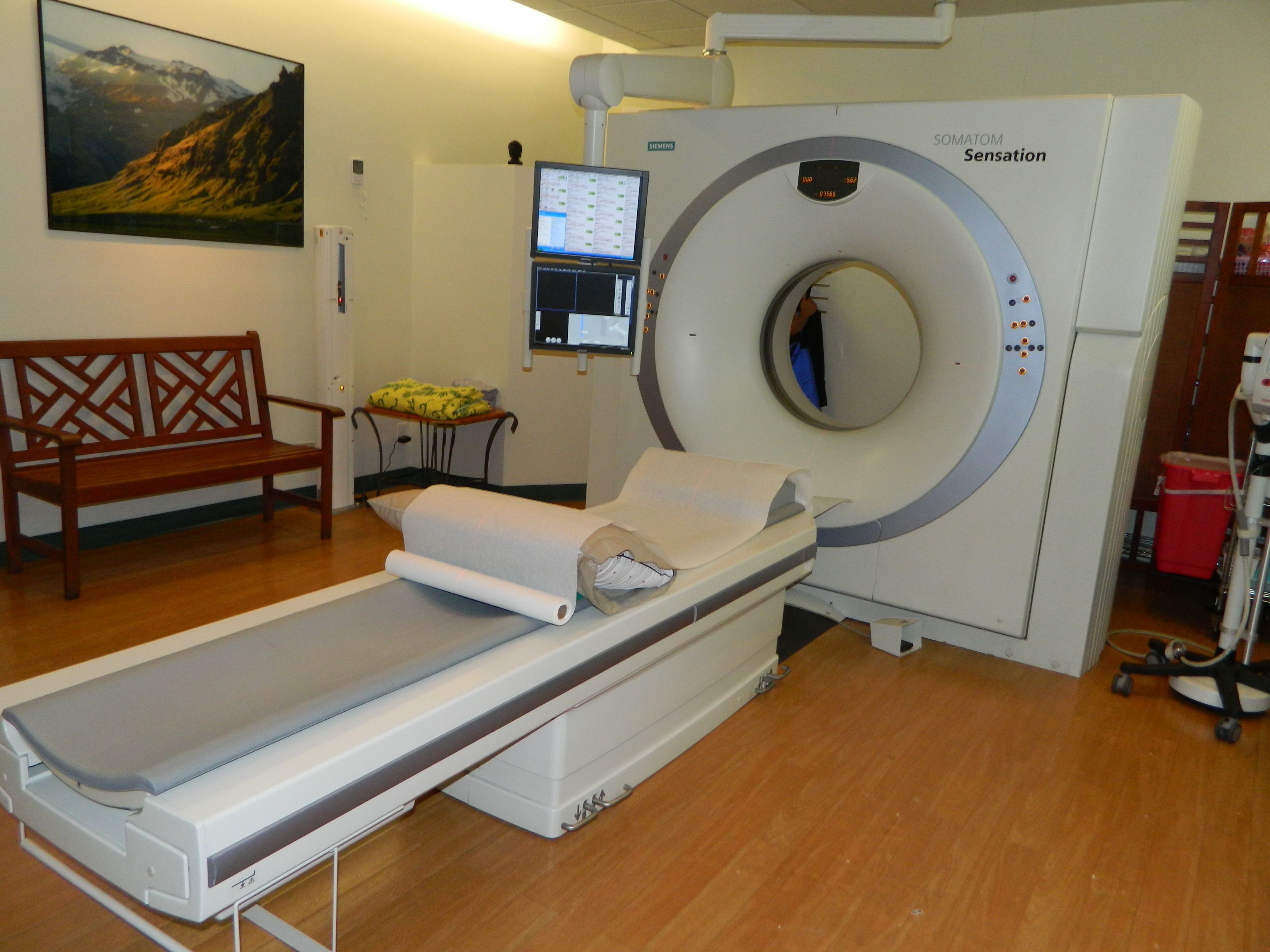
 Computed Tomography (CT/ CAT scan)
With the advent of multi-slice (or multi-detector) technology, Computed Tomography (CT) is now a routine diagnostic imaging modality that is versatile and reliably answers many referring doctor’s clinical questions.
A CT scanner uses an X-ray beam that revolves around a patient to produce a cross sectional image known as a “slice”. Modern CT scanners use multi-slice technology, which means that many fine slices may be performed of the patient as they enter the CT scanner in a single rotation, with the processing of the data resulting in reconstruction of the image from any angle. This means that 3-D images may be produced and assist in the formulation of a diagnosis.
Computed Tomography is of particular use in the diagnosis of chest, abdominal and pelvic disorders, including the bowel and lungs, as well as in the production of images of arteries (known as an angiogram). A CT angiogram can therefore be used to diagnose a brain aneurysm, blockage (stenosis) of an artery, including the arteries of the heart and of the legs. Importantly, the diagnosis of a life threatening blood clot to the lungs, known as a pulmonary embolus, is reliably made. Multi-slice CT is also a suitable modality for evaluating the spine.
Patients unable to have an MRI scan may undergo either a CT and/or Ultrasound in order to address their medical complaint.
As a CT uses rapid X-ray technology, it can also be used for sophisticated spine injection procedures, as well as guiding a needle deep into body parts for a specific medical purpose, such as a biopsy or drainage.
Referring doctors are welcome to discuss with our radiologist the imaging needs of their patients and whether CT is suitable for their patient’s medical condition.
